

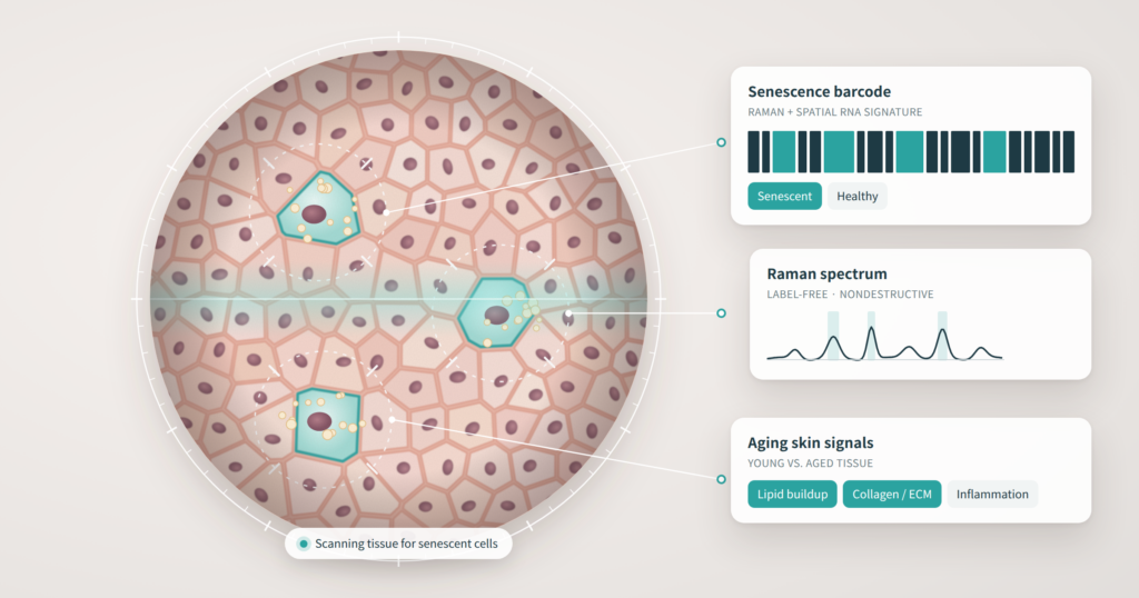
What if a dermatologist could see which cells in your skin had quietly stopped working, without cutting out a single sample? A team from MIT, Massachusetts General Hospital, and Harvard Medical School has taken a meaningful step in that direction. In a study published in Nature Aging on September 21, 2026, the researchers describe a noninvasive way to identify senescent cells, often called “zombie cells,” using light instead of a scalpel.
The work is early and was done in mice. But for anyone following the science behind “anti-aging” skincare, it is one of the more substantive developments of the year.
What Are Zombie Cells?
Senescent cells are cells that have permanently stopped dividing but refuse to die. The trigger is often DNA damage. Once senescent, these cells change shape, rewire their metabolism, and shift their gene expression.
In young tissue, the immune system clears them out efficiently. With age, that cleanup slows down, and senescent cells accumulate. The MIT team notes that this buildup may contribute to sagging skin, muscle weakness, and chronic conditions like osteoarthritis and type 2 diabetes.
It is worth stressing that senescence is not purely harmful. It plays important roles in embryonic development and tissue repair, which is exactly why researchers want to identify these cells precisely rather than simply wipe them out.
The Problem With Today’s Markers
Scientists already have biomarkers for senescence, most notably the cell cycle proteins p16 and p21. The catch: detecting them requires processing that destroys the cells being studied.
That is a serious limitation. You cannot watch the same cells over time, and you certainly cannot use a destructive test as a routine clinical screen on a patient’s skin.
Two Views of the Same Cell
The researchers turned to Raman microscopy, an optical technique that shines near-infrared or visible light on tissue and reads the scattered light to reveal its chemical makeup. Crucially, it leaves the tissue intact.
On its own, a Raman spectrum tells you about chemistry, not biology. So the team paired it with spatial RNA sequencing, which maps where genes are switched on within a tissue. Measuring both in the same cells gave them a much richer profile: gene activity, location, and biochemical composition, all at once. The published method is called RamanOmics.
They applied this combined approach to skin and lung tissue from 2-month-old and 26-month-old mice.
What Changed in Aging Skin
Several findings stand out for dermatology:
- Lipid buildup: Older skin and lung cells both showed increased lipid synthesis and lipid accumulation. What this means for cell function is still unknown.
- Collagen and matrix remodeling: In senescent skin cells, pathways tied to collagen and extracellular matrix remodeling were significantly affected.
- Contractile pathways: Skin cells also showed changes in pathways associated with muscle contraction.
- Tissue-specific signatures: Aged lung tissue, by contrast, showed more activity in immune and inflammatory genes, a reminder that senescence does not look the same everywhere.
The collagen and matrix finding is the one to watch. Collagen loss and matrix breakdown are central to visible skin aging, and much of the cosmeceutical and aesthetic market is built around them. A tool that links those changes to specific senescent cells, in intact tissue, would give researchers a far more direct way to test whether a treatment is actually working.
From Barcode to Diagnostic
By combining the most informative Raman peaks with the most informative gene signatures, the team built a senescence “barcode.” The practical payoff: in the future, a device might only need to scan a handful of specific Raman bands to flag senescent cells, rather than running a full molecular workup.
The long-term vision includes endoscope-style tools that could detect senescence inside the body. For skin, the more obvious application is an optical probe applied to the surface.
The current bottleneck is speed. Analyzing a tissue sample of roughly one square millimeter takes about 30 hours. The team is now building a faster imaging system designed to pick out just the barcode bands across larger areas.
Separating Signal From Headline
The press coverage describes this as an “AI-powered” barcode, and this is where a careful reader should slow down. The real innovation here is the pairing of two measurement techniques and the computational work of identifying which features best predict senescence. That is legitimate, data-driven science. But the public summary says little about the specific algorithms involved, and the “AI” framing risks overshadowing what is genuinely new: nondestructive, spatially resolved detection of senescent cells.
A few other points to keep in view:
- This is a mouse study. The researchers are working on adapting it to human tissue, but human validation has not yet been reported.
- No clinical test exists yet. There is no device a patient or clinician can use today, and the current imaging time rules out routine use.
- Skin of color is an open question. Optical techniques applied to human skin must contend with melanin, which is known to interfere with many light-based measurements. Any human version of this barcode will need to be validated across the full range of skin tones before it can be trusted clinically.
The Bigger Picture
Senescence has become a buzzword in skincare marketing, with products promising to target “zombie cells” long before anyone could reliably see those cells in living skin. Research like this, backed by the NIH Cellular Senescence Network, points toward a future where such claims could actually be measured.
That is the real significance. A noninvasive senescence readout would not just aid diagnosis of age-related disease. It could become an objective endpoint for evaluating senolytic drugs, procedures, and topical products, separating treatments that change skin biology from those that simply claim to. The technology is still in early stages, but the direction is clear, and it is one dermatology should be watching closely.
—
Source: MIT News: “Unmasking ‘zombie cells’ in aging tissue with an AI-powered barcode”